This document was ed by and they confirmed that they have the permission to share it. If you are author or own the copyright of this book, please report to us by using this report form. Report r6l17
Overview 4q3b3c
& View Bedside & Central Monitoring System as PDF for free.
More details 26j3b
- Words: 1,361
- Pages: 37
BEDSIDE & CENTRAL MONITORING SYSTEM
BY: VISHAL UPADHYAY ICE-B 110115098
INTRODUCTION TO PATIENT MONITORING SYSTEMS ●
Used for monitoring physiological signals including Electrocardiograph (ECG), Respiration, Invasive and NonInvasive Blood Pressure, Oxygen Saturation in Human Blood, Body Temperature and other Gases etc.
●
Multiple sensors and electrodes used for receiving physiological signals like ECG Electrodes, SpO2 Finger Sensor, Blood Pressure Cuff and Temperature Probe to measure the physiological signals.
●
During treatment, it is highly important to continuously monitor the vital physiological signs of the patient. Therefore, patient monitoring systems has always been occupying a very important position in the field of medical devices.
CLASSES OF PATIENT MONITORING SYSTEM ●
Single-Parameters Monitoring Systems
●
Multi-Parameter Patient Monitoring Systems
●
The single parameter monitoring system is available for measuring blood pressure of a human body, ECG(Electrocardiograph) monitor, SpO2 (Oxygen Saturation inBlood) monitor etc.
●
A multi-parameter Patient Monitoring System (PMS) is used for multiple critical physiological signs of the patient to transmit the vital information like Electrocardiograph,Respiration Rate, Blood pressure etc.
●
Most diseases of the heart and of the circulatory system,referred to as cardiovascular diseases, strike without warning and prompt treatment is required if death is to be averted. Such treatment is best provided in a specialized area of hospital referred to as “Intensive Care Unit”(ICU).
●
These specialized hospital units provide constant observation of the subject, constant monitoring of the subject’s physiological condition and provide immediate emergency treatment whenever it is required.
THREE IMPORTANT INTENSIVE CARE UNITS ●
Coronary intensive care units Used for treatment of diseases of the heart such as the heart attacks.
●
Stroke intensive care Units Used for treatment of diseases of the circulatory system such as stroke.
●
Pulmonary intensive care unit Used for treatment of respiratory diseases
Who requires patient monitoring? ●
Patients with unstable physiologic regulatory systems; Example: a patient whose respiratory system is suppressed by a drug overdose or anesthesia.
●
Patients with a suspected life-threatening condition; Example: a patient who has findings indicating an acute myocardial infarction (heart attack).
●
Patients at high risk of developing a life-threatening condition; Example: patients immediately post open-heart surgery, or a premature infant whose heart and lungs are not fully developed.
●
Patients in a critical physiological state; Example: patients with multiple trauma or septic shock.
●
Mother and baby during the labor and delivery process.
Why patient monitoring?
Data Acquisition and Signal Processing
General Block Diagram of Medical Instrumentation System
Block Diagram of Bedside Monitoring System
Interfacing Analog Signals to Microprocessors
Display ●
Digital Storage Oscilloscope(DSO) are used.
●
Advanced trigger.
●
High Speed Sampling.
●
Data not lost.
Nursing Flowsheet
Present Parameters in Patient Monitoring System • ECG 3/5/10 leads • Respiration • Invasive Blood Pressure (IBP) • Non Invasive Blood Pressure (NIBP) • Dual Temperature • Pulse Oxy Meter (SpO2)
Common Patient Monitoring Systems
Cardiac(ECG) Monitoring ●
Cardiac monitoring is used to continuously monitor the heart’s rhythm.
●
A normal heartbeat is called a sinus rhythm.
●
It can be represented by six distinct waves, identified in the image to the left by the letters P, Q, R, S, T, and U.
Cardiac(ECG) Monitoring
Cardiac(ECG) Monitoring
Driven right leg circuit ●
A Driven Right Leg Circuit or DRL circuit is an electric circuit that is often added to biological signal amplifiers to reduce Common-mode interference.
●
Biological signal amplifiers such as ECG (Electrocardiogram) EEG (Electroencephalogram) or EMG circuits measure very small electrical signals emitted by the body, often as small as several micro-volts.
●
The patient's body can also act as an antenna which picks up electromagnetic interference, especially 50/60 Hz noise from electrical power lines.
●
Right Leg Driver circuitry is used to eliminate interference noise by actively canceling the interference.
Driven right leg circuit
Measurement of Heart Rate
●
ECG is sampled every 2ms.
●
High amplitude component & artifacts are reduced by Slew Rate Limiter.
●
High Frequency component & DC offset voltage is removed with help of filters.
●
Filtered ECG signal is ed through QRS matched filter.
●
Detector recognizes QRS complex that has occurred since last heart beat.
●
If this value exceeds threshold value, a heart beat is counted.
Hemodynamic monitoring ●
Blood pressure (BP) and blood flow are monitored by a hemodynamic monitor and can be done in two ways:
●
Blood pressure cuff (Non-invasive)
●
With an intra-arterial catheter.(Invasive)
Non-invasive Blood Pressure Monitoring ●
Manual or automated devices
●
Different Methods of measurement:
●
Oscillometric (Electronic Pressure sensor)
●
The Rheographic Method
●
Auscultatory (Korotkoff sounds)
Oscillometric Method
The Rheographic Method
Direct Arterial Blood Pressure Measurement (Invasive Method)
Direct Arterial Blood Pressure Measurement (Invasive Method)
Pulse Oximeter
(Respiratory monitoring) ●
Sensors used to continuously measure the Oxygen saturation (SPO2) of haemoglobin in blood. It displays the percentage of blood that is loaded with oxygen.
Principle of Pulse Oximeter ●
●
Principle of pulse oximetry is based on the differential absorption characteristics of oxygenated and the deoxygenated hemoglobin. Oxygenated hemoglobin absorbs more infrared light and allows more red light to through. Whereas Deoxygenated hemoglobin absorbs more red light and allowing more infrared light to through.
What’s inside the Sensor? ●
Each pulse oximeter sensor probe contains two light emitting diode: one emitting red light and the other emitting near infrared light.
●
It also has a photo-detector.
●
The photo-detector measures the intensity of transmitted light at each wavelength.
●
And using the differences in the reading the blood oxygen content is calculated.
●
The probe is placed on a suitable part of the body, usually a fingertip or ear lobe.
Methods for Monitoring Oxygen Saturation in Blood ●
Transmission Method
●
Reflectance Method
Transmission Method ●
●
●
●
●
●
Transmitter (LED) & the receiver(photo-detector) are placed on opposite side of the finger. Finger will be placed between the LED’s & the photo-detector. When the finger is placed a part of the light will be absorbed by the finger and some part will reach the photo detector. Now with each heart beat there will be increase in volume of blood flow this will result in more light getting absorbed by the finger so less light reaches the photo-detector. Hence if we see the waveform of received light signal it will consist of peaks in between heart beats and trough (bottom) at each heartbeat. This difference between the trough & the peak value is the reflection value due to blood flow at heart beat
Reflectance Method ●
LED & photo-detector are placed on the same side i.e. next to each other.
●
In the reflective method there will be some fixed light reflection back to the sensor due to finger.
●
With each heart-beat there will be an increase in blood volume in the finger this will result in more light reflection back to the sensor.
●
Hence if we see the waveform of the received light signal it will consist of peaks at each heartbeat.
●
A fixed low value reading is there in between the heart beats this value can be considered as constant reflection and this difference of the peak subtracted from the constant reflection value is the reflection value due to blood flow at heart beat.
●
Body temperature monitoring: An adhesive pad containing a thermoelectric transducer is used to monitor body temperature.
●
Blood glucose monitoring: In this technique for testing the amount of glucose (sugar) in the blood, a small amount of blood (usually a drop), is extracted from from the fingertip and placed on the end of a coated strip, called a testing strip. The strip changes color according to the amount of glucose in the blood. Modern hospitals often allow the results of the testing to be transmitted wirelessly to the patient’s EMR.
●
Childbirth monitoring: Also called fetal monitoring, it can be done by fetoscope, ultrasound, or electrode and pressure catheter.
General Considerations In Monitoring System ●
Inaccessibility of the Signal Source
●
Variability of Physiological Signals
●
Interference among Physiological System
●
Transducer Interface Problems
●
High Possibility of Artifacts
●
Safe levels of Applied Energy
●
Patient Safety Considerations
●
Reliability Aspects
●
Human factor Considerations
●
Government Regulations
THANK YOU
Related Documents 171j1w

November 2019 41
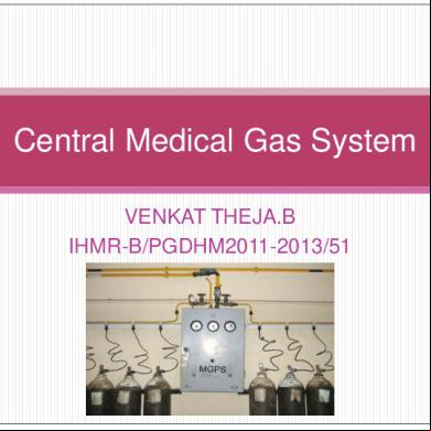
Central Medical Gas System 6cu1b
December 2020 0
Central Nervous System 4mf6c
April 2023 0
Bedside Nurse bi15
November 2019 54
Iot Garbage Monitoring System 3j5h5m
April 2022 0
Visual Monitoring System e2b1m
October 2022 0More Documents from "EPS" 583p1f
November 2019 41
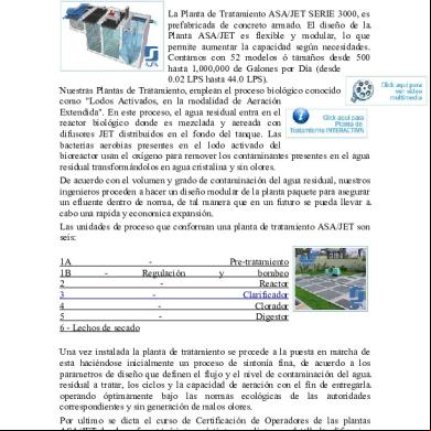
Planta De Tratamiento De Aguas Residuales Comercial 6b1a5
January 2023 0
2c82t
April 2020 16
2c82t
September 2021 0
Evaluative Statement 2uz5y
November 2022 0